
Epicondylitis (Grade 1-2)
Authors: Francesco Porta1
- 1Pistoia, Italy
- Publisher
- Tiss’You
- Published
- Document ID
- #OP2FP
Version
Original English text of the study.
Source document
On this page
Key findings
- VNR assessment evidenced a decrease in pain in all follow-ups with a major benefit after second infiltration.
- Ultrasound evaluations showed reductions in tendon edema, changes in lesion characteristics, and improved echogenicity, with better clarity.
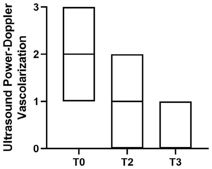
- Power-Doppler evaluation evidenced a decrease in vascularization, indicating a reduction in inflammation and restoration of the tendon’s normal morphology.
- Some patients displayed limited improvements, with residual edema or partially visible lesions.
- No adverse events or complications were observed during the treatment process.
- patients
- 13
- injections of 1 ml (day 0 and day 15)
- 2
- follow-up
- 45 days
- hydrolyzed collagen peptides
- 5 mg/ml
- Indication
- Epicondylitis, Grade 1-2.
- Medical device
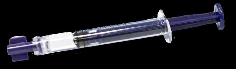
- Hydrolyzed Collagen 5 mg/1 ml. Medical device based on low molecular weight collagen peptides (LWPs). It is a ready-to-use injectable solution for the structural strengthening of connective tissues.
- Manufacturer
- Tiss’You
- Patients
- 13 patients, 34-72 years
- Investigator
- Francesco Porta
- Location
- Pistoia, Italy
- Treatment
- Two injections of 1 ml of 5 mg/ml hydrolyzed collagen peptides: at day 0 and at day 15 (secondary treatment).
- Follow-up
- Baseline, 7 days, 15 days and 45 days.
- Report
- PMCF (Post-Market Clinical Follow-up) Report #OP2FP, 20 June 2023.
Epicondylitis, commonly known as tennis elbow or golfer’s elbow, is a prevalent condition characterized by inflammation and microtears in the tendons attaching to the lateral or medial epicondyle of the elbow. It affects a significant number of individuals, particularly those engaged in repetitive wrist and arm movements. Current treatment options for epicondylitis include rest, physical therapy, nonsteroidal anti-inflammatory drugs (NSAIDs), corticosteroid injections, and in severe cases, surgery. However, these approaches have limitations, such as short-term relief, potential side effects, and variable efficacy.
Hydrolyzed collagen peptides offer a promising alternative for the treatment of epicondylitis. These peptides, derived from collagen protein, possess unique regenerative properties that can aid in tissue repair and restoration. Local injection of collagen peptides directly into the affected tendon area allows for targeted delivery and enhanced bioavailability. Once administered, the peptides interact with the damaged tendon, promoting cellular proliferation, migration, and the synthesis of extracellular matrix components. Moreover, collagen peptides exhibit anti-inflammatory effects, reducing local inflammation and relieving pain associated with epicondylitis.
Thirteen patients, ranging in age from 34 to 72 years, presented with Epicondylitis graded at levels 1 and 2. These patients underwent a treatment regimen involving two injections of 1 ml of Low Molecular Weight Collagen Peptides (LWPs), administered at days 0 and 15. To assess the treatment’s effectiveness, patients underwent evaluations at key time points, including baseline, 7 days, 15 days, and 45 days post-treatment. Pain levels were quantified using the Visual Numeric Rating (VNR) scale, while the assessment of treatment response included Power-Doppler ultrasound analysis and qualitative analysis of relevant parameters.
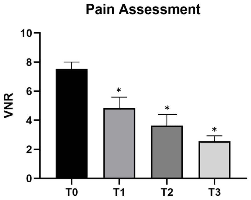
VNR assessment evidenced a decrease in pain in all follow-ups with a major benefit after second infiltration.
The collective analysis of the pre-treatment and post-treatment ultrasound evaluations reveals varying outcomes in response to the treatment. Overall, there were instances of positive progress, including reductions in tendon edema, changes in lesion characteristics, and improved echogenicity, with better clarity. Furthermore, Power-Doppler evaluation evidenced a decrease in vascularization, indicating a reduction in inflammation and restoration of the tendon’s normal morphology. These findings indicate a positive response to the treatment, supporting the notion of improved tissue healing and reduced inflammation.
However, it is worth noting that some patients displayed limited improvements, with residual edema or partially visible lesions, indicating the need for further intervention or alternative treatment approaches to achieve more substantial results.
It is important to consider these collective findings alongside other clinical information to comprehensively evaluate the overall effectiveness of the treatment approach. Importantly, no adverse events or complications were observed during the treatment process, highlighting the safety of the intervention.
Citation
Porta F. Epicondylitis, Grade 1-2. PMCF Report #OP2FP. Tiss’You; 20 June 2023.